Saturday 31 October 2015

Low Cost Heart Bypass Surgery in India - Best Heart Surgery Hospital India

Heart Bypass Surgery in India

Coronary artery bypass grafting (CABG) or Heart Bypass is a type of surgery that improves blood flow to the heart. Surgeons use CABG to treat people who have severe coronary heart disease (CHD). Since Longer life expectancy has led to an increased incidence of cardiovascular disease and consequently to an increasing number of primary and secondary coronary artery bypass grafting (CABG) operations in the elderly. A secondary Bypass surgery is called a Redo Bypass Surgery. Redo Bypass is a relatively difficult procedure and requires an extensive experience by the heart surgeon an extensive to perform the heart bypass surgery.
Why is it required?
CHD is a disease in which high cholesterol levels would lead to formation of a waxy substance called plaque inside the coronary arteries. These arteries supply oxygen-rich blood to your heart. Over time, plaque can harden or rupture (break open). Hardened plaque narrows the coronary arteries and reduces the flow of oxygen-rich blood to the heart. This can cause chest pain or discomfort called Angina.
If the plaque ruptures, a blood clot can form on its surface. A large blood clot can mostly or completely block blood flow through a coronary artery. This is the most common cause of a heart attack. Over time, ruptured plaque also hardens and narrows the coronary arteries.
A Redo-CABG is required in cases where the Coronary arteries narrow down again due to the existing CHD leading to the formation of plaque again in the Coronary arteries.
What is done during CABG/Redo CABG?
During CABG/Redo CABG, a healthy artery or vein from the body is connected, or grafted, to the blocked coronary artery by the surgeon. The heart surgeon ensures that the grafted artery or vein bypasses (that is, goes around) the blocked portion of the coronary artery. This creates a new path for oxygen-rich blood to flow to the heart muscle.
The Goals of CABG/Redo CABG include:
  Improving your quality of life and reducing angina and other CHD symptoms
  Allowing you to resume a more active lifestyle
  Improving the pumping action of your heart if it has been damaged by a heart attack
  Lowering the risk of a heart attack (in some patients, such as those who have diabetes)
  Improving your chance of survival

Friday 30 October 2015

Ventricular Septal Defect in Children - Ventricular Septal Defect Surgery in India

Ventricular Septal Defect(VSD)
A small hole allows little amount of blood to go across the hole. A large hole gets a lot more of blood across the hole. This extra amount of blood coming to the right side of the heart immediately gets transmitted to the lungs from where it goes to the left side of the heart. So, it is really the left side if the heart, the left upper and lower chambers which get to see this extra amount of blood!
VSD when large has to be closed by the 3rd to 6th month of life.  The decision to close the
VSD has to be made well in time to give family enough time to accommodate to the fact that
heart surgery has to be done on their child and that apart from the emotions of the decision
(how so ever safe it might be, parents will take time to decide and accept), there are
financial (or insurance clearance issues also).
  1. VSD with Coarctation
  1. VSD, large with a pneumonia
  1. Large VSD with Severe Malnutrition
What are the ventricles ? What is the Ventricular septum ?
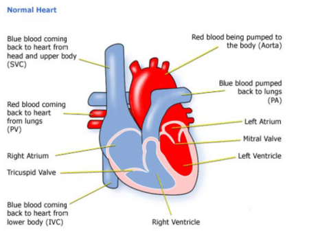
The heart has 2 pumps; pumping blood is the main function of the heart; the 2 pumps are separated by a wall called the ventricular septum. The right pump receives the blue blood which has to go to the lungs for picking up oxygen. This pump has to push blood into its vicinity (heart and lungs being close by within the chest); so the normal pressure in this pump is 30-35 mmHg.
The Left Pump pushes the red blood into the lungs; it has to ensure blood reaches all the parts of the body so the pressure here is equivalent to the blood pressure (upper number of BP i.e. Systolic).
What is a Ventricular septal defect (VSD) ?
A defect in the wall which separates the ventricles is called a VSD. The high pressure in the Left pump as compared to the Right ensures that the blood flows from the left to the right pump (therefore is called a L to R shunt). This implies red (left sided) blood gets into the blue (right sided) blood; this implies that in this condition the patient is not going to be blue.
How common is it?
It is one of the most common defects of the heart. Infact it is the most common defect.
Can it be detected before birth ? 
Yes, it can be detected before and at birth. Often, it will detected on an ultrasound. But on fetal echocardiogram there is a better chance of it being picked up. The way this hole is noticed, is by seeing abnormal flow across the septum. But, during fetal life the pressure between the 2 pumps is the same. So, there is not much flow across the hole and that makes it difficult to pick up.
How does a small hole differ from a large hole?
If this blood amount is large, the left side of the heart gets dilated and volume overloaded. This can be seen on an X-ray as enlarged heart.  This reverses once the extra blood flow stops.
If the amount of blood is large, there will also be an increase in pressure in the right side of heart and in the lungs. While with the Small VSD the volume will be so small the left heart won’t enlarge. And neither will the pressure in the right heart go up.

Is VSD closure an emergency ?
Usually VSD closure is an elective procedure done in a planned manner, but, ocassioanlly
may turn out to be emergency in some circumstances:
A ventricular septal defect usually is diagnosed after a baby is born.

Doctors may choose to monitor and observe children who have ventricular septal defects (VSDs) but don't have symptoms of heart failure. This means regular checkups and tests to see whether the defect closes on its own or gets smaller.
More than half of VSDs eventually close, usually by the time a child is in preschool. Your child's doctor will let you know how often your child should be checked. Checkups may range from once a month to once every 1 or 2 years.
When treatment for a VSD is required, options include extra nutrition and surgery to close the VSD.
The doctor may recommend surgery if your child's VSD:

Some infants who have VSDs don't grow and develop or gain weight as they should. These infants usually:
Doctors usually recommend extra nutrition or special feedings for these infants. These feedings are high-calorie formulas or breast milk supplements that give babies extra nourishment.
In some cases, tube feeding is needed. Food is given through a small tube that's placed through the nose and into the stomach. Tube feeding can add to or take the place of bottle feeding. This treatment usually is temporary because a VSD that causes symptoms will likely need surgery.

Most doctors recommend surgery to close large VSDs that are causing symptoms or haven't closed by the time children are 1 year old. Surgery may be needed earlier if:
Rarely, medium-sized VSDs that are causing enlarged heart chambers are treated with surgery after infancy. However, most VSDs that need surgery are repaired in the first year of life.
Diagnosis
 
  • Shortness of breath,
  • Fast or heavy breathing,
  • Sweating,
  • Tiredness while feeding, or
  • Poor weight gain.
  • Is large
  • Is causing symptoms
  • Is medium-sized and is causing enlarged heart chambers
  • Affects the aortic valve
Extra Nutrition
  • Have large VSDs
  • Are born prematurely
  • Tire easily during feeding
  • The child fails to gain weight
  • Medicines are needed to control the symptoms of heart failure

Wednesday 28 October 2015

Low Cost Pediatric Cardiac Surgery at Best Cardiac Hospital in India


Paediatric Heart Surgery in India with the Best Paediatric Heart surgeons in India is what you would want if your little one is suffering from any heart ailment. MyMedOpinion affiliate Paediatric heart surgeons in India are among the top paediatric heart specialists and offer best in paediatric heart care. Affordable Pediatric Heart Surgery in India at Best Cardiac Hospitals with Top Pediatric Cardiac Surgeons in India with MyMedOpinion.

Sometimes heart surgery in children is required for repairing defects in the heart which a child might have born with (known as congenital heart defects) and heart diseases which he gets after birth, which require surgery. Pediatric Cardiac Surgery deals with operative procedures in the newborn and unborn children and youngsters suffering from cardiac dysfunctions, structural, functional and rhythm-related issues of the heart also.

In the normal heart, electrical impulses arise from an area of specialized cells called the sinus node, which is the heart's normal pacemaker. The sinus node is located in the right atrium, the upper right chamber of the heart . After leaving the sinus node, an impulse spreads across the upper heart chambers (right and left atria) and reaches the atrioventricular (AV) node located near the center of the heart between the atria and the lower chambers, or ventricles, and then to conduction fibers that spread across the ventricles, which are the heart's main pumping chambers. 

As the impulse moves along the conduction fibers, cardiac muscle cells are stimulated and contract, producing a heartbeat.Pediatric Cardiac Surgery often deals with heart problems in children, the cause of congenital heart diseases which are common heart ailments among new born children and involves deficiencies like structural defects, congenital arrythmias and cardiomyopathies, which result in different kinds of abnormalities related to the heart. Congenital heart disease is a cause of improper growth of the heart or blood vessels before birth.


Heart Treatment in India for Major Heart Ailments in Children
The most common cardiac defects in children are the following:

• Atrial Septal Defect
• Ventricular Septal Defect
• Fallot's Tetralogy
• Valvular defects

Atrial Septal Defect
An atrial septal defect (ASD) — sometimes referred to as a hole in the heart — is a type of congenital heart defect in which there is an abnormal opening in the dividing wall between the upper filling chambers of the heart (the atria). In most cases ASDs are diagnosed and treated successfully with few or no complications. When an atrial septal defect is present, blood flows through the hole primarily from the left atrium to the right atrium.
This shunting increases the blood volume in the right atrium which means more blood flows through the lungs than would normally. If left untreated, atrial septal defect may cause problems in adulthood. These problems may include pulmonary hypertension (which is high blood pressure in the lungs), congestive heart failure(weakening of the heart muscle), atrial arrhythmias (which are abnormal rhythms or beating of the heart) and an increased risk of stroke.

Ventricular Septal Defect - VSD have a hole in the wall of their heart between their right ventricle and left ventricle (the two lower chambers, where the blood leaves the heart). Normally, blood cannot pass between the ventricles. But when there is a hole between the sides of the heart, some oxygen-rich blood leaks from the left ventricle into the right ventricle and goes back to the lungs. The hole may be small and cause no symptoms, or it may be large and cause serious problems with blood flow. If the hole is large, too much blood will be pumped to the lungs, leading to congestive heart failure. Also, the heart will have to work harder to pump blood to the body. As a result of the extra work, the heart can get bigger.

There are two main techniques to correct the disorder:
Intra-Cardiac Technique - is an open heart surgery under general anesthesia , whereby the child is attached to an external heart-lung machine. This heart lung machine performs circulatory and respiratory function during surgery. The heart is directly accessed and the defect is sutured.

Trans- Catheter Technique - this is a minimally invasive technique through which surgical instruments are passed through the catheter inserted in the femoral artery. The catheter is slowly guided up towards the point of defect to close it.
Both the procedures are quite successful. Which one is best for your child can be ascertained by the paediatric cardiac surgeon in India after the examination of the child.

Fallot's Tetralogy
Fallot was a doctor who spotted this particular type of heart defect. Tetralogy means fourfold – there are four defects found together. These four problems are: 1. Pulmonary stenosis - Pulmonary means ‘of the lungs’. Stenosis means narrowing. Pulmonary stenosis is a narrowing at or below the pulmonary valve. 2. Ventricular septal defect -Ventricular means ‘of the ventricles’ – the wall between the right and left sides of the heart . 3. Over-riding aorta -The entrance to the aorta, which should only take red (oxygenated) blood around the body, lies over the VSD, allowing the right ventricle to pump some blue blood directly into it. 4. Right ventricular hypertrophy -The right ventricle becomes thickened as it forces blood into the narrowed pulmonary artery.

The heart surgeons perform two surgeries to correct Fallot of Tetralogy heart defect in the child Blalock- Taussig's operation is a palliative procedure performed in smaller infants to increase blood flow to lungs and to allow the child to grow big enough to withstand the corrective surgery. A connection is made between right subclavian artery and pulmonary artery to pass more oxygenated blood to the latter. This relieves the cyanosis to a great extent.

The total corrective surgery is performed in children within 2 years of age. VSD is closed with a patch and the narrowed pulmonary valve is opened. The outcome of surgery is favourable and most children lead a healthy life after the surgery with minimum restrictions

Double valve repair and replacement
Valves are openings between two chambers and also between a chamber and artery which allows unidirectional flow of blood. The patency of these valves is very important for normal flow of blood. The function of mitral valve (between left atrium and left ventricle) and the aortic valve (between left ventricle and aorta) is very important. If these valves get narrowed, the amount of blood passing from left atrium to left ventricle (mitral valve) or from left ventricle to aorta (aortic valve) is drastically reduced. In this case the heart will pump blood harder to push it through the narrowed valves. But the blood will tend to go upwards back into the pulmonary vein and finally to the lungs. 

This will cause excess blood in the lungs leading to congestion. At the same time very less blood will come out into the aorta from the left ventricle which will result in reduced blood and oxygen supply to all the body parts.

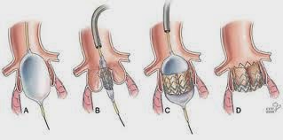
Double valve repair and replacement procedure aims at correcting or replacing these both these damaged valves (aortic & mitral together) with new functional valves. This is done through the open heart surgery. The patient is put under general anaesthesia and connected to the heart lung machine. This machine takes over the pumping, circulatory, and respiratory functions of the heart and lung till the surgical procedure is going on. The valves are accessed by cutting open the rib cage and accessing the heart directly. The old valves are sliced from their attachments and new valves are put in their place. 

The new valves may be obtained from a cadaveric donor, or an animal (pig) or it may be made of a nonreactive inert material. The patient needs to be given anticoagulants or immunosuppressive to clotting of blood or rejection of organ. The prognosis of this surgery is good. It is many a time a life-saving and life extending surgery.


Radio frequency catheter ablation is a procedure that is performed to correct a disturbance in heart rhythm (cardiac arrhythmia or irregular heartbeat).
It`s most often used to treat supraventricular tachyarrhythmias which are rapid, uncoordinated heartbeats starting in the heart`s upper chambers (atria) or middle region (AV node or the very beginning portion of the heart`s electrical system). In cardiology, RF ablation is used to correct:
• recurrent atrial flutter.
• atrial fibrillation (AF).
• supraventricular tachycardia (SVT).
• some types of ventricular arrhythmia.


About MyMedOpinion.com

MyMedOpinion affiliated  Best hospitals in India provide an medical opinion from experienced surgeons and the treatment cost includes companion stay  , surgeon fee, medicines and consumables, nursing care, patient's food and airport pick up & drop etc. etc. We offer free, no obligation assistance to international patients to find world class medical treatment in India. We offer support and services to facilitate the care you require. We can help you find the best hospital in India

Send us a Medical Report to Get FREE Medical Opinion from India's Top Doctors

MyMedOpinion.com  is the hub of Surgeons and specialists for major diseases. We give you a chance to speak to our doctors and discuss your health issues directly. 
Email Us: Free@mymedopinion.com  Call Us: +91-7503537224

Sunday 25 October 2015

Heart Valve Repair or Replacement Surgery in India - Best Heart Hospital India

Heart Valve Repair or Replacement Surgery 

Blood is pumped through your heart in only one direction. Heart valves play a key role in this one-way blood flow, opening and closing with each heartbeat. Pressure changes on either side of the valves cause them to open their flap-like "doors" (called cusps or leaflets) at just the right time, then close tightly to prevent a backflow of blood.
There are 4 valves in the heart:
  • Tricuspid valve
  • Pulmonary valve
  • Mitral valve
  • Aortic valve

Nearly all of these operations are done to repair or replace the mitral or aortic valves. These valves are on the left side of the heart, which works harder than the right. They control the flow of oxygen-rich blood from the lungs to the rest of the body.

Valve Disease
If valve damage is mild, doctors may be able to treat it with medicines. If damage to the valve is severe, surgery to repair or replace the valve may be needed.

What is valve repair?
Valve repair can usually be done on congenital valve defects (defects you are born with) and has a good success record with treating mitral valve defects.
Here are some procedures surgeons may use to repair a valve:
  • Commissurotomy, which is used for narrowed valves, where the leaflets are thickened and perhaps stuck together. The surgeon opens the valve by cutting the points where the leaflets meet.
  • Valvuloplasty, which strengthens the leaflets to provide more support and to let the valve close tightly. This support comes from a ring-like device that surgeons attach around the outside of the valve opening.
  • Reshaping, where the surgeon cuts out a section of a leaflet. Once the leaflet is sewn back together, the valve can close properly.
  • Decalcification, which removes calcium buildup from the leaflets. Once the calcium is removed, the leaflets can close properly.
  • Repair of structural support, which replaces or shortens the cords that give the valves support (these cords are called the chordae tendineae and the papillary muscles). When the cords are the right length, the valve can close properly.
  • Patching, where the surgeon covers holes or tears in the leaflets with a tissue patch.
What is valve replacement?
Severe valve damage means that the valve will need to be replaced. Valve replacement is most often used to treat aortic valves and severely damaged mitral valves. It is also used to treat any valve disease that is life-threatening. Sometimes, more than one valve may be damaged in the heart, so patients may need more than one repair or replacement.
There are 2 kinds of valves used for valve replacement:
  • Mechanical valves, which are usually made from materials such as plastic, carbon, or metal. Mechanical valves are strong, and they last a long time. Because blood tends to stick to mechanical valves and create blood clots, patients with these valves will need to take blood-thinning medicines for the rest of their lives.
  • Biological valves, which are made from animal tissue (called a xenograft) or taken from the human tissue of a donated heart (called an allograft or homograft). Sometimes, a patient's own tissue can be used for valve replacement (called an autograft). Patients with biological valves usually do not need to take blood-thinning medicines. These valves are not as strong as mechanical valves, though, and they may need to be replaced every 10 years or so. Biological valves break down even faster in children and young adults, so these valves are used most often in elderly patients.
You and your doctor will decide which type of valve is best for you.
During valve repair or replacement surgery, the breastbone is divided, the heart is stopped, and blood is sent through a heart-lung machine. Because the heart or the aorta must be opened, heart valve surgery is open heart surgery.

What to Expect
The operation will usually be scheduled at a time that is best for you and your surgeon, except in urgent cases. As the date of your surgery gets closer, be sure to tell your surgeon and cardiologist about any changes in your health. If you have a cold or the flu, this can lead to infections that may affect your recovery. Be aware of fever, chills, coughing, or a runny nose. Tell the doctor if you have any of these symptoms.

Also, remind your cardiologist and surgeon about all of the medicines you are taking, especially any over-the-counter medicines such as aspirin or those that might contain aspirin. You should make a list of the medicines and bring it with you to the hospital.
It is always best to get complete instructions from your cardiologist and surgeon about the procedure, but here are some basics you can expect when you have valve repair or replacement surgery.

Before the Hospital Stay
Most patients are admitted to the hospital the day before surgery or, in some cases, on the morning of surgery.
The night before surgery, you will be asked to bathe to reduce the amount of germs on your skin. After you are admitted to the hospital, the area to be operated on will be washed, scrubbed with antiseptic, and, if needed, shaved.
A medicine (anesthetic) will make you sleep during the operation. This is called "anesthesia." Because anesthesia is safest on an empty stomach, you will be asked not to eat or drink after midnight the night before surgery. If you do eat or drink anything after midnight, it is important that you tell your anesthesiologist and surgeon.
If you smoke, you should stop at least 2 weeks before your surgery. Smoking before surgery can lead to problems with blood clotting and breathing.

Day of Surgery
Before surgery, you may have an electrocardiogram (ECG or EKG), blood tests, urine tests, and a chest x-ray to give your surgeon the latest information about your health. You will be given something to help you relax (a mild tranquilizer) before you are taken into the operating room.

Small metal disks called electrodes will be attached to your chest. These electrodes are connected to an electrocardiogram machine, which will monitor your heart's rhythm and electrical activity. You will receive a local anesthetic to numb the area where a plastic tube (called a line) will be inserted in an artery in your wrist. An intravenous (IV) line will be inserted in a vein. The IV line will be used to give you the anesthesia before and during the operation.

After you are completely asleep, a tube will be inserted down your windpipe and connected to a machine called a respirator, which will take over your breathing. Another tube will be inserted through your nose and down your throat, into your stomach. This tube will stop liquid and air from collecting in your stomach, so you will not feel sick and bloated when you wake up. A thin tube called a catheter will be inserted into your bladder to collect any urine produced during the operation.

A heart-lung machine is used for all valve repair or replacement surgeries. This will keep oxygen-rich blood flowing through your body while your heart is stopped. A perfusion technologist or blood-flow specialist operates the heart-lung machine. Before you are hooked up to this machine, a blood-thinning medicine called an anticoagulant will be given to prevent your blood from clotting. The surgical team is led by the cardiovascular surgeon and includes other assisting surgeons, an anesthesiologist, and surgical nurses.

After you are hooked up to the heart-lung machine, your heart is stopped and cooled. Next, a cut is made into the heart or aorta, depending on which valve is being repaired or replaced. Once the surgeon has finished the repair or replacement, the heart is then started again, and you are disconnected from the heart-lung machine.\

The surgery can take anywhere from 2 to 4 hours or more, depending on the number of valves that need to be repaired or replaced.

Recovery Time
You can expect to stay in the hospital for about a week, including at least 1 to 3 days in the Intensive Care Unit (ICU).

Recovery after valve surgery may take a long time, depending on how healthy you were before the operation. You will have to rest and limit your activities. Your doctor may want you to begin an exercise program or to join a cardiac rehabilitation program.
If you have an office job, you can usually go back to work in 4 to 6 weeks. Those who have more physically demanding jobs may need to wait longer.


Minimally invasive heart valve surgery is a technique that uses smaller incisions to repair or replace heart valves. This means there is less pain. Minimally invasive surgery also reduces the length of the hospital stay and the recovery time.
Minimally invasive valve surgery can only be done in certain patients. This type of surgery cannot be done in patients
  • With severe valve damage
  • Who need more than one valve repaired or replaced
  • Who have clogged arteries (atherosclerosis)
  • Who are obese
In some cases, minimally invasive valve surgery can be done using a robot. Robotic surgery does not require a large incision in the chest. It is not available at all hospitals, and patients with severe valve damage cannot have the procedure. The Texas Heart Institute has a robot.

With robotic surgery, the surgeon has a control console, a side cart with 3 robotic arms, a special vision system, and instruments. A computer translates the surgeon's natural hand and wrist movements made on the control console to instruments that have been placed inside the patient through small incisions. The robot's controls can read even the tiniest of movements the surgeon makes.
Robotic surgery can reduce the time it takes to do valve surgery, as well as shorten the hospital stay and recovery time. 

Cardiac Surgery at World Best Hospitals in India

Robotic Heart Surgery in India 

Popularly known as the Bypass Surgery, Cardiac bypass implanting a healthy or working blood vessel from any other part of the body to bypass the blocked vessel. The heart surgeons in India are highly experienced to perform a CABG or heart bypass surgery. India offers great value proposition to patients looking for affordable Heart surgery or Cardiac surgery abroad as not only the cost of cardiac bypass surgery in India is very low, the facilities and infrastructure is most modern and is counted among the best heart hospitals in the world. 


The blockages in the arteries grow with the deposition of fat. This process is called as Artherosclerosis. These blockages obstruct the free flow of the blood to the various parts of the heart leading to severe chest pain (known as angina) or a heart attack in the later stages.Today Cardiology treatment in India has come up as a suitable option in order to get rid of any of the heart defects as the cost in India of any of the treatments is the best and that too at rates which are absolutely affordable. Because of these benefits of choosing in India, any of the treatments, many foreigners have come down here in order to solve their trouble of heart diseases. 


When repair is not an option, replacing your damaged heart valve may be the most effective treatment for your condition. Also known as Heart Valve Replacement, Aortic Valve Replacement, is a cardiac surgery procedure in:-


  • Ring Annuloplasty - In this procedure the surgeon repairs the ring-like part around the valve by stitching a ring of metal, cloth, or tissue around the valve.
  • Valve repair - In this one or more leaflets of the valve is trimmed, shaped, or rebuild. The leaflets are the flaps that are responsible for opening and closing of the valve.


Pediatric Cardiothoracic Surgery 
Pediatric cardiothoracic Surgery is a kind of heart surgery that is performed children for repairing heart defects which occur at the time of birth which are known as congenital heart diseases. Pediatric Cardiac Surgery is necessary for the well-being of the child.
This surgery is done to take care of various diseases in children such as Artrial Septal Defect (ASD), Patent Ductus Arteriosus and Total correction or Four Abnormality correction (TOF). 

Heart Surgery Procedures performed with Excellent Outcomes:-

  • CABG - Minimally Invasive Valve & Coronary Heart Bypass Techniques.
  • Off-pump Coronary Artery Bypass (OPCAB)
  • Mitral & Aortic Valve repair & restoration surgery
  • Aortic dissection surgery
  • Transmyocardial laser revascularization
  • Congenital and Pediatric heart surgery
  • Heart failure surgery- Ventricular restoration and Ventricular assist Devices
  • Carotid endarterectomy
  • Peripheral vascular procedures
  • Atrial Fibrillation & Arrhythmia surgery 


The most advanced technology for minimally invasive heart surgery is now available in India. Robotic da Vinci Surgical System combines superior 3D visualization along with greatly enhanced dexterity, precision and control in an intuitive, ergonomic interface with breakthrough surgical capabilities.In Robotic Cardiac Surgery, robotic assist the heart surgeon in performing surgery through tiny incisions. The surgeon has to sit in an operating room where he controls the instruments while looking at 3D images from the camera which is inside the patient. Various procedures can be performed in this case such as mitral valve replacement or repair, correction of HOCM etc. Use of Robotic Surgery in Cardiology has benefits as it involves less pain, scarring and risk that are otherwise involved. The surgeon can perform surgery through tiny openings in the chest, cracking the breastbone and spreading the ribs. 

Minimally Invasive Surgery
Much modern cardiothoracic surgery is performed with the help of highly advanced machines that are operated externally by a surgeon. The main advantage to these machines is that they can operate through very small incisions, and this dramatically shortens the recovery time of a patient. The most advanced machines are even able to react intelligently to changes in the patient’s body. For instance, some machines can adapt to the beating of a heart, moving their arms in synch with it, so that to the surgeon operating the machine it appears as if the heart is stationary.