Tuesday 24 November 2015

Affordable Coronary Bypass Heart Surgery (CABG) in India

Coronary bypass surgery performed in world class hospitals in India is a procedure to allow blood to flow to your heart muscle despite blocked arteries. Coronary bypass surgery uses a healthy blood vessel taken from your leg, arm, chest or abdomen and connects it to the other arteries in your heart so that blood is bypassed around the diseased or blocked area. After a coronary bypass surgery, normal blood flow is restored. Coronary bypass surgery is just one option to treat heart disease. 


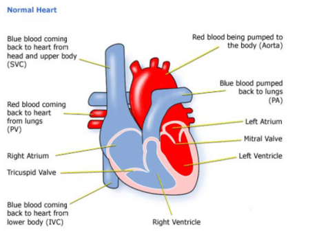
Just like all the other organs in your body, your heart needs blood and oxygen to do its job. Coronary arteries snake across the surface of your heart, delivering a constant supply of blood and oxygen to the heart muscle. When one or more of these arteries become narrowed or blocked, blood and oxygen are reduced and heart muscle is damaged. Coronary bypass surgery can minimize this damage. 


Today Cardiology treatment in India has come up as a suitable option in order to get rid of any of the heart defects as the cost in India of any of the treatments is the best and that too at rates which are absolutely affordable. Because of these benefits of choosing in India, any of the treatments, many foreigners have come down here in order to solve their trouble of heart diseases.
Who Should Consider Coronary Artery Bypass Graft Surgery?
  • People diagnosed with arterial blockage or heart damage are recommended with the Coronary Artery Bypass Graft Surgery.
  • People suffering from severe chest pain or angina due to the arterial blockage are recommended with the Coronary Artery Bypass Graft Surgery.
  • People suffering from complicated conditions such as diabetes & high blood pressure are recommended the Coronary Artery Bypass Graft Surgery to reduce the risk of heart attack.

Procedure for Coronary Bypass Surgery

During a Coronary Artery Bypass Graft Surgery (CABG), the blood flow is re-routed around the clogged artery by detaching a long segment of an artery from the chest wall, arms or leg veins. Thereafter, the new artery is grafted to the clogged area of the coronary artery. Through the newly attached channel, blood gets unhindered route to flow to the heart muscles. This procedure is known as Coronary Artery Bypass Surgery. Depending upon the number of blocked coronary arteries, a patient may undergo more than one bypass graft.
What to Expect After Coronary Artery Bypass Graft Surgery?
After surgery, there will be a short stay (1 to 2 days if there are no complications) in the intensive care unit (ICU). In the ICU, you will likely have:
·         Continuous monitoring of his or her heart activity.
·         A tube to temporarily help with breathing.
·         A stomach tube, to remove stomach secretions until the person starts eating again.
·         A tube (catheter) to drain the bladder and measure urine output.
·         Tubes connected to veins in the arms (intravenous, or IV, lines) through which fluids, nutrition, and medicine can be given.
·         An arterial line to measure blood pressure.
·         Chest tubes, to drain the chest cavity of fluid and blood (which is temporary and normal) after surgery.
Benefits of Coronary Artery Bypass Graft Surgery
Some of the potential benefits of Coronary Bypass Heart Surgery (CABG) include :
  • Lower risk of stroke
  • Lower death rate
  • Less need for transfusion
  • Less heart rhythm problems
  • Less injury to the heart
FAQ's - Coronary Artery Bypass Graft Surgery in India

What is the cost of Coronary Artery Bypass Graft Surgery?
Coronary Artery Bypass Graft Surgery is amongst the lowest in the world. The Coronary Artery Bypass Graft Surgery is about 20% of the cost in the USA. The low Coronary Artery Bypass Graft Surgery is without any compromise on quality or success rate

Are cardiac surgeons in India well qualified to perform heart surgeries?
Teaching hospitals in India are of a high standard. Further, many cardiac surgeons and cardiologists from India have had further education/training abroad at top class medical schools and hospitals. The result is a very high level of academic excellence amongst cardiologists and cardiac surgeons in India.

How experienced are cardiac surgeons in India at handling complex heart surgeries?
The translation of academic excellence into outstanding medical results happens only with practice and experience. Indian doctors acquire a great amount of experience over a very short period of time because of the large number of patients requiring cardiac treatment in India.

Do surgeons perform advanced heart surgeries in India?
Cardiac surgeons in India are experts at performing advanced procedures like Heart Transplants, Robotic Cardiac Procedures, Totally Endoscopic Coronary Artery Bypass Surgery (TECAB), Minimally Invasive Direct Coronary Artery Bypass Grafting (MIDCAB), Off-Pump Coronary Artery Bypass Grafting (OPCAB), complex mitral valve repairs, etc.

What success rates can one expect in India from cardiology hospitals?
It is quite amazing that Indian hospitals are able to offer a combination of extremely low cost cardiac treatment in India along with extremely high success rates as well. In fact, leading hospitals for cardiac treatment in India have success rates in excess of 98%, which is absolutely world-class.

Get a free of cost second opinion from renowned surgeons within 24 hours.

For further information or free consultation, we request you to fill in inquiry form on the website and send medical reports to free@mymedopinion.com. MyMedOpinion assures you that all arrangements for examinations, surgeries, and post-operative recuperation will be ably taken care of you

Get the MyMedOpinion Advantage

MyMedOpinion help by its expertise and speed and quality of response. From arranging opinion from India's best cancer hospitals, hospital appointment bookings, travel and hotel accommodation, we manage our patient’s needs efficiently.
·         Quick responses… Within 24-hours
·         World class results for  treatment
·         Cancer specialists with great qualifications and experience

·         Assistance to Plan your Travel and Apply for India visa.

Friday 6 November 2015

Pacemaker implantation Surgery in India | Cost of Pacemaker Surgery in India

Heart rhythm problems (arrhythmia) occur when the electrical impulses produced by your heart that coordinate heartbeat do not function properly, causing your heart to beat too quickly, too slowly, or irregularly. Age increases the probability of experiencing an arrhythmia. It can occur in people who do not have heart disease. Some heart arrhythmias are harmless, though some types, such as ventricular tachycardia (fast heart rates), are serious and even life threatening.

Pacemakers represent one of the earliest and most successful nonpharmacological therapy for arrhythmias. Millions of pacemakers have been implanted since the very first pacemaker was implanted. Drugs are no longer used except in the very acute setting before implantation of a temporary or permanent pacemaker.

A cardiac pacemaker is a device that is used to regulate the heart rate.
If you have been found to have a heartbeat that is too slow, a pacemaker can be implanted in the body to take over the function. This small electronic device automatically monitors and regulates the heartbeat, by transmitting electrical impulses to stimulate the heart when it is beating too slowly. A pacemaker consists of a pacing lead and a pulse generator. Single chamber pacemakers have only a single lead while dual chamber pacemakers have two leads with one lead in the atrium and the other in the ventricle. Dual chamber pacemakers are more physiological but more expensive.

Types of pacemakers

Single chamber pacemakers are pacing systems that use one lead in either the right atrium or the right ventricle of your heart.

A single lead in the right atrium is commonly used in conditions where the normal pacemaker of the heart is not working adequately, such as in the case of sick sinus syndrome. Atrial pacing is used when the sinus node is sending out signals that are too slow or irregular. However, to use this method of pacing, the rest of the heart's normal conduction system must be functioning normally.

More commonly, the single lead is placed in the right ventricle to help correct a slow or irregular heart beat. This is most often the case when the electrical flow is slowed or blocked in the region of the atrio-ventricular (A-V) node and the normal impulses from the atria cannot reach the ventricle. This would result in too slow a heart beat. The pacemaker system would keep the heart beating at a steady rate

Dual chamberpacemakers are pacemaker systems that use a lead in the right atrium as well as the right ventricle (figure 6). This type of pacing most closely mimics the heart's normal conduction pattern by pacing sequentially from atria to ventricle thus maximizing the heart's pumping ability. By having a lead in both the atria and ventricle the pulse generator is able to continuously regulate the heart's electrical activity in both chambers. These are the most commonly used pacemakers at the present time.

Commonly asked questions about pacemakers

Will I need to make any lifestyle changes after my pacemaker is implanted?
There are no significant lifestyle changes that you will need to make as a result of having a pacemaker implanted. Most patients resume their normal activities soon after implantation. Specific issues or concerns should be addressed with your pacemaker physician or nurse.
How often will I need to have my pacemaker checked?
Your pacemaker system will need to be evaluated by your pacemaker physician, nurse, or your local cardiologist's office at least twice yearly. A special computer called a programmer is used to perform a comprehensive evaluation of your pacemaker system. The programmer has a wand (like a computer mouse) that is used to communicate with the pacemaker. The wand is placed on your chest directly over the pulse generator and a radio wave signal is used to send and receive information from the pulse generator. Changes in the pacemaker settings can be done via this method as well. A complete assessment of the pacemaker's sensing and pacing functions, battery life, and diagnostic information is obtained, which enables your pacemaker physician/nurse to fine tune your care.
How is the battery changed?
The battery that is used to power your pulse generator is tightly sealed within the metal shell of the device. Therefore, when the battery's energy is depleted a whole new pulse generator must be implanted. The skin over the pulse generator site is numbed up with local anesthetic. You may also receive a light sedative through a intravenous line to help you relax. A new incision is made in the skin and the pacemaker pocket is opened. The pulse generator is removed and lead(s) disconnected. At this time the lead(s) are hooked to a special analyzer that evaluates the lead(s) for any evidence of potential malfunction. A new pulse generator is then attached to the lead(s) and the system is reimplanted in the same pocket. The incision is sutured (sewn) together and a small dressing applied. Most patients can go home the same day as their procedure.
Can I use a microwave?
Microwave ovens will not interfere with the proper functioning of your pacemaker. You can use a microwave oven without concern.
Can I use a cell phone?
It is possible that a cellular phone might interfere with the normal functioning of your pacemaker. The interaction is temporary, however, and will only affect the pacemaker during the time that your cellular phone is close to your pacemaker. To avoid this potential interference, it is recommended that you hold the cellular phone on the opposite side of your body away from the pacemaker. You should also not store your cellular phone in your breast pocket.You should always try to maintain a distance of at least 6 inches between your cellular phone and your pacemaker system.
Do I have to take any precautions at the airport?
If you were to walk through the metal detector at the airport, it will not harm you nor your pacemaker. However, because the pacemaker is encased in a metal shell, it is possible that the pacemaker may set off the security alarm. To avoid this problem, it is generally recommended that you show your pacemaker identification card to the security agent and inform him/her that you have an implanted pacemaker system. They should let you pass around the metal detector. If the airport security wants to scan you with the "hand wand", they can everywhere except over the device. This information also pertains to any metal detector such as at a courthouse or federal building.


Thursday 5 November 2015

Open Heart Surgery Hospital India - Heart bypass surgery cost in India


Open Heart Surgery is one of the most complicated and complex surgeries in the world, and only the most expert and skilled medical professionals should undertake it.  India, we have some of the most renowned and skilled surgeons in the world, and we provide the top of the line infrastructure and medical devices, to ensure that the patients are in the best hands possible, and have all the necessary supporting devices and infrastructure. Indian Hospitals offer all the necessary amenities and services that one would require to get back to normal health, no matter what their medical issue is.

The Hospitals are  complete service center for heart and other organs related surgeries and medical attention. Our diagnosis culture is one of the best in the world, and it provides critical and complete information necessary for the medical personnel to understand and undertake the kind of medical treatment that would be perfect for the current medical issue. We have the necessary and state of the art amenities and support system, which make it the best option for people of all nationalities when it comes to open heart surgeries and other critical and complex surgeries.

Indications for Open Heart Bypass Surgery:

http://www.indiahealthtour.com/images/homepage/li.jpgMajor diet changes, smoking cessation and exercise have not improved the    coronary artery disease. 

http://www.indiahealthtour.com/images/homepage/li.jpgAngioplasty is unsuccessful, not appropriate or the artery is becoming blocked again after angioplasty. 

http://www.indiahealthtour.com/images/homepage/li.jpgSevere chest pain with activity. 

http://www.indiahealthtour.com/images/homepage/li.jpgTesting shows severe disease in the left main coronary artery. 

http://www.indiahealthtour.com/images/homepage/li.jpgTesting shows severe disease in multiple arteries. 

http://www.indiahealthtour.com/images/homepage/li.jpgStents did not work, could not be placed or are needed again. 

http://www.indiahealthtour.com/images/homepage/li.jpgThe left ventricle is not working properly due to coronary artery disease. 

http://www.indiahealthtour.com/images/homepage/li.jpgThere is an imminent risk of heart damage. 


Preparing for Open Heart Bypass Surgery:

Many cardiac surgeons order extensive testing before proceeding with a CABG to determine which arteries are obstructed and the severity of the blockage. An angiogram is one outpatient test that uses x-rays to determine the severity of coronary disease. 

A stress test, electrocardiogram (EKG) and blood tests are usually done in advance of the surgery. Blood tests may be repeated immediately prior to surgery to determine if the patient is likely to bleed during surgery, along with their general health. 

A surgeon may have very specific instructions for a patient scheduled for bypass surgery. These instructions may include changes in prescription medications, diet and drinking and smoking habits


The cost of heart surgery, yet there are no hard and fast rules as to exactly find the net cost of the heart surgery. For example – the surgeon’s fee may vary with the medical center chosen. For instance, the charges may vary if a bigger hospital facility is chosen as compared to a smaller one, even if the surgeon is same in both the set-ups. Likewise, the severity of heart condition alone or together with the associated risk factors and medical conditions such as obesity or high blood sugar may alter the treatment plan and thus would alter the cost of the heart surgery.

 In countries such as Russia and countries such as Kenya, Tanzania, Uganda and other east-african countries, where the condition of medical facilities is quite grim, medical tourism is a boon for these countries. With rise of medical tourism and ever improving healthcare facilities in India, more and more people from these countries are heading to India for various heart surgery procedures.

Get a free of cost second opinion from renowned surgeons within 24 hours.
For further information or free consultation, we request you to fill in inquiry form on the website and send medical reports to free@mymedopinion.com. MyMedOpinion assures you that all arrangements for examinations, surgeries, and post-operative recuperation will be ably taken care of you

Get the MyMedOpinion Advantage
MyMedOpinion help by its expertise and speed and quality of response. From arranging opinion from India's best cancer hospitals, hospital appointment bookings, travel and hotel accommodation, we manage our patient’s needs efficiently.
·Quick responses… Within 24-hours
·World class results for  treatment
·Cancer specialists with great qualifications and experience
·Assistance to Plan your Travel and Apply for India visa.

Types of Valve Replacement Surgery - Valve Replacement Surgery in India

Valvular heart disease is a form of heart disease that occurs when one or more of the heart’s four valves don’t function properly. Valve replacement surgery is a solution for patients whose valves are too fragile, scarred, or otherwise damaged to repair.

Reasons for Replacement

The valves of the heart are responsible for allowing nutrient-rich blood to flow to the chambers of your heart. Like a locked gate, each valve is supposed to close completely after ushering in the blood flow. Diseased heart valves aren’t always able to perform the job as well as they should.

Stenosis, or a narrowing of the passageway, causes a less-than-normal amount of blood to flow to the heart, causing the muscle to work harder. Leaky valves can also pose a problem. Instead of closing tightly, a valve may remain slightly open, letting blood flow backwards. Signs of valvular heart disease can include:
  • fatigue
  • dizziness or lightheadedness
  • shortness of breath
  • chest pain
  • fluid retention, especially in the lower limbs
Heart valve repair is also a solution for valve disease, but in some people, the damage is too far advanced and a total replacement of the affected valve is required.

Types of Replacement Valves

Mechanical and biologic valves are used to replace faulty valves. Mechanical valves are artificial components that fulfill the same job as a natural heart valve. They’re created from carbon and polyester materials that are well-tolerated by the human body, and can last between 10 and 20 years, according to the National Institutes of Health (NIH). However, one of the risks associated with mechanical valves is blood clots. Recipients of mechanical heart valves are required to take blood-thinning medication for the rest of their lives to reduce their risk of stroke.

Biologic valves, also called bioprosthetic valves, are created from human or animal tissue. Three types of biologic heart valves exist:
  • human: called allografts or homografts, tissue is taken from a donor heart
  • porcine: made from pig tissue, the valves can be implanted with or without a frame called a stent
  • bovine: crafted from cow tissue, bovine valves connect to your heart with silicone rubber
Biologic valves don’t increase your risk of developing blood clots. Therefore, you most likely won’t need to commit to a lifetime of anti-clotting medication. The lifespan of a bioprosthetic isn’t as long as a mechanical valve, explains the Mayo Clinic, and may require replacement at a future date. Your age, overall health, age, and ability to take anti-coagulant medications, as well as the extent of the disease, will help determine which type of valve you receive.

Types of  Valve Replacement Surgery

Aortic Valve Replacement

The aortic valve is located on the left side of the heart and serves as an outflow valve. Its job is to allow blood to leave the left ventricle (the heart’s main pumping chamber) and close so that blood doesn’t leak back into the ventricle. Surgery on the aortic valve may be required due to a congenital (from birth) defect or disease that causes the valve to narrow (stenosis) or leak (regurgitation).

Mitral Valve Replacement

The mitral valve is located on the left side of the heart. It serves as an inflow valve. Its job is to allow blood from the left atrium to flow into the heart’s main pumping chamber, the left ventricle. Surgery may be required if the valve doesn’t fully open or completely close. When the valve is too narrow, blood may have difficulty entering and can back up, causing pressure in the lungs. When the valve doesn’t close properly, blood can leak back into the lungs. This can be due to a congenital defect, infection, or a degenerative disease (a worsening over time due to age or normal wear and tear).

Double Valve Replacement

Double valve replacement is replacement of both the mitral and the aortic valve, or the left side of the heart. This type of surgery is not as common as the others and the mortality rate is slightly higher. 

Pulmonary Valve Replacement

The pulmonary valve separates the pulmonary artery—which carries blood to the lungs for oxidation—and the right ventricle (one of the heart’s chambers). Its job is to allow blood to flow from the heart to the lungs, via the pulmonary artery. The need for pulmonary valve replacement is usually due to stenosis, which restricts blood flow. Stenosis may be caused by a congenital defect, infection, or carcinoid syndrome.     


Heart valve replacement surgery is performed under general anesthesia with techniques that are either conventional or minimally invasive. Conventional surgery requires a large incision spanning from your neck to your navel. Less invasive measures can reduce the length of incision and also reduce your risk of infection.

For a surgeon to successfully remove the diseased valve and replace it with a new one, your heart must be still. You’ll be placed on a bypass machine that keeps blood circulating through your body and your lungs functioning during surgery. The surgeon will make incisions into your aorta, through which the valves will be removed and replaced. Cleveland Clinic reports a 1.8 percent risk of death associated with valve replacement surgery.

Recovery


The majority of heart valve replacement recipients remain in the hospital for approximately five to seven days. If your surgery was minimally invasive, you may be able to go home earlier. Medical staff will offer pain medication as needed and continuously monitor your blood pressure, breathing, and heart function during the first few days after a heart valve replacement.Full recovery may take a few weeks or up to several months, depending on your rate of healing


Saturday 31 October 2015

Low Cost Heart Bypass Surgery in India - Best Heart Surgery Hospital India

Heart Bypass Surgery in India

Coronary artery bypass grafting (CABG) or Heart Bypass is a type of surgery that improves blood flow to the heart. Surgeons use CABG to treat people who have severe coronary heart disease (CHD). Since Longer life expectancy has led to an increased incidence of cardiovascular disease and consequently to an increasing number of primary and secondary coronary artery bypass grafting (CABG) operations in the elderly. A secondary Bypass surgery is called a Redo Bypass Surgery. Redo Bypass is a relatively difficult procedure and requires an extensive experience by the heart surgeon an extensive to perform the heart bypass surgery.
Why is it required?
CHD is a disease in which high cholesterol levels would lead to formation of a waxy substance called plaque inside the coronary arteries. These arteries supply oxygen-rich blood to your heart. Over time, plaque can harden or rupture (break open). Hardened plaque narrows the coronary arteries and reduces the flow of oxygen-rich blood to the heart. This can cause chest pain or discomfort called Angina.
If the plaque ruptures, a blood clot can form on its surface. A large blood clot can mostly or completely block blood flow through a coronary artery. This is the most common cause of a heart attack. Over time, ruptured plaque also hardens and narrows the coronary arteries.
A Redo-CABG is required in cases where the Coronary arteries narrow down again due to the existing CHD leading to the formation of plaque again in the Coronary arteries.
What is done during CABG/Redo CABG?
During CABG/Redo CABG, a healthy artery or vein from the body is connected, or grafted, to the blocked coronary artery by the surgeon. The heart surgeon ensures that the grafted artery or vein bypasses (that is, goes around) the blocked portion of the coronary artery. This creates a new path for oxygen-rich blood to flow to the heart muscle.
The Goals of CABG/Redo CABG include:
  Improving your quality of life and reducing angina and other CHD symptoms
  Allowing you to resume a more active lifestyle
  Improving the pumping action of your heart if it has been damaged by a heart attack
  Lowering the risk of a heart attack (in some patients, such as those who have diabetes)
  Improving your chance of survival

Friday 30 October 2015

Ventricular Septal Defect in Children - Ventricular Septal Defect Surgery in India

Ventricular Septal Defect(VSD)
A small hole allows little amount of blood to go across the hole. A large hole gets a lot more of blood across the hole. This extra amount of blood coming to the right side of the heart immediately gets transmitted to the lungs from where it goes to the left side of the heart. So, it is really the left side if the heart, the left upper and lower chambers which get to see this extra amount of blood!
VSD when large has to be closed by the 3rd to 6th month of life.  The decision to close the
VSD has to be made well in time to give family enough time to accommodate to the fact that
heart surgery has to be done on their child and that apart from the emotions of the decision
(how so ever safe it might be, parents will take time to decide and accept), there are
financial (or insurance clearance issues also).
  1. VSD with Coarctation
  1. VSD, large with a pneumonia
  1. Large VSD with Severe Malnutrition
What are the ventricles ? What is the Ventricular septum ?
The heart has 2 pumps; pumping blood is the main function of the heart; the 2 pumps are separated by a wall called the ventricular septum. The right pump receives the blue blood which has to go to the lungs for picking up oxygen. This pump has to push blood into its vicinity (heart and lungs being close by within the chest); so the normal pressure in this pump is 30-35 mmHg.
The Left Pump pushes the red blood into the lungs; it has to ensure blood reaches all the parts of the body so the pressure here is equivalent to the blood pressure (upper number of BP i.e. Systolic).
What is a Ventricular septal defect (VSD) ?
A defect in the wall which separates the ventricles is called a VSD. The high pressure in the Left pump as compared to the Right ensures that the blood flows from the left to the right pump (therefore is called a L to R shunt). This implies red (left sided) blood gets into the blue (right sided) blood; this implies that in this condition the patient is not going to be blue.
How common is it?
It is one of the most common defects of the heart. Infact it is the most common defect.
Can it be detected before birth ? 
Yes, it can be detected before and at birth. Often, it will detected on an ultrasound. But on fetal echocardiogram there is a better chance of it being picked up. The way this hole is noticed, is by seeing abnormal flow across the septum. But, during fetal life the pressure between the 2 pumps is the same. So, there is not much flow across the hole and that makes it difficult to pick up.
How does a small hole differ from a large hole?
If this blood amount is large, the left side of the heart gets dilated and volume overloaded. This can be seen on an X-ray as enlarged heart.  This reverses once the extra blood flow stops.
If the amount of blood is large, there will also be an increase in pressure in the right side of heart and in the lungs. While with the Small VSD the volume will be so small the left heart won’t enlarge. And neither will the pressure in the right heart go up.

Is VSD closure an emergency ?
Usually VSD closure is an elective procedure done in a planned manner, but, ocassioanlly
may turn out to be emergency in some circumstances:
A ventricular septal defect usually is diagnosed after a baby is born.

Doctors may choose to monitor and observe children who have ventricular septal defects (VSDs) but don't have symptoms of heart failure. This means regular checkups and tests to see whether the defect closes on its own or gets smaller.
More than half of VSDs eventually close, usually by the time a child is in preschool. Your child's doctor will let you know how often your child should be checked. Checkups may range from once a month to once every 1 or 2 years.
When treatment for a VSD is required, options include extra nutrition and surgery to close the VSD.
The doctor may recommend surgery if your child's VSD:

Some infants who have VSDs don't grow and develop or gain weight as they should. These infants usually:
Doctors usually recommend extra nutrition or special feedings for these infants. These feedings are high-calorie formulas or breast milk supplements that give babies extra nourishment.
In some cases, tube feeding is needed. Food is given through a small tube that's placed through the nose and into the stomach. Tube feeding can add to or take the place of bottle feeding. This treatment usually is temporary because a VSD that causes symptoms will likely need surgery.

Most doctors recommend surgery to close large VSDs that are causing symptoms or haven't closed by the time children are 1 year old. Surgery may be needed earlier if:
Rarely, medium-sized VSDs that are causing enlarged heart chambers are treated with surgery after infancy. However, most VSDs that need surgery are repaired in the first year of life.
Diagnosis
 
  • Shortness of breath,
  • Fast or heavy breathing,
  • Sweating,
  • Tiredness while feeding, or
  • Poor weight gain.
  • Is large
  • Is causing symptoms
  • Is medium-sized and is causing enlarged heart chambers
  • Affects the aortic valve
Extra Nutrition
  • Have large VSDs
  • Are born prematurely
  • Tire easily during feeding
  • The child fails to gain weight
  • Medicines are needed to control the symptoms of heart failure

Wednesday 28 October 2015

Low Cost Pediatric Cardiac Surgery at Best Cardiac Hospital in India


Paediatric Heart Surgery in India with the Best Paediatric Heart surgeons in India is what you would want if your little one is suffering from any heart ailment. MyMedOpinion affiliate Paediatric heart surgeons in India are among the top paediatric heart specialists and offer best in paediatric heart care. Affordable Pediatric Heart Surgery in India at Best Cardiac Hospitals with Top Pediatric Cardiac Surgeons in India with MyMedOpinion.

Sometimes heart surgery in children is required for repairing defects in the heart which a child might have born with (known as congenital heart defects) and heart diseases which he gets after birth, which require surgery. Pediatric Cardiac Surgery deals with operative procedures in the newborn and unborn children and youngsters suffering from cardiac dysfunctions, structural, functional and rhythm-related issues of the heart also.

In the normal heart, electrical impulses arise from an area of specialized cells called the sinus node, which is the heart's normal pacemaker. The sinus node is located in the right atrium, the upper right chamber of the heart . After leaving the sinus node, an impulse spreads across the upper heart chambers (right and left atria) and reaches the atrioventricular (AV) node located near the center of the heart between the atria and the lower chambers, or ventricles, and then to conduction fibers that spread across the ventricles, which are the heart's main pumping chambers. 

As the impulse moves along the conduction fibers, cardiac muscle cells are stimulated and contract, producing a heartbeat.Pediatric Cardiac Surgery often deals with heart problems in children, the cause of congenital heart diseases which are common heart ailments among new born children and involves deficiencies like structural defects, congenital arrythmias and cardiomyopathies, which result in different kinds of abnormalities related to the heart. Congenital heart disease is a cause of improper growth of the heart or blood vessels before birth.


Heart Treatment in India for Major Heart Ailments in Children
The most common cardiac defects in children are the following:

• Atrial Septal Defect
• Ventricular Septal Defect
• Fallot's Tetralogy
• Valvular defects

Atrial Septal Defect
An atrial septal defect (ASD) — sometimes referred to as a hole in the heart — is a type of congenital heart defect in which there is an abnormal opening in the dividing wall between the upper filling chambers of the heart (the atria). In most cases ASDs are diagnosed and treated successfully with few or no complications. When an atrial septal defect is present, blood flows through the hole primarily from the left atrium to the right atrium.
This shunting increases the blood volume in the right atrium which means more blood flows through the lungs than would normally. If left untreated, atrial septal defect may cause problems in adulthood. These problems may include pulmonary hypertension (which is high blood pressure in the lungs), congestive heart failure(weakening of the heart muscle), atrial arrhythmias (which are abnormal rhythms or beating of the heart) and an increased risk of stroke.

Ventricular Septal Defect - VSD have a hole in the wall of their heart between their right ventricle and left ventricle (the two lower chambers, where the blood leaves the heart). Normally, blood cannot pass between the ventricles. But when there is a hole between the sides of the heart, some oxygen-rich blood leaks from the left ventricle into the right ventricle and goes back to the lungs. The hole may be small and cause no symptoms, or it may be large and cause serious problems with blood flow. If the hole is large, too much blood will be pumped to the lungs, leading to congestive heart failure. Also, the heart will have to work harder to pump blood to the body. As a result of the extra work, the heart can get bigger.

There are two main techniques to correct the disorder:
Intra-Cardiac Technique - is an open heart surgery under general anesthesia , whereby the child is attached to an external heart-lung machine. This heart lung machine performs circulatory and respiratory function during surgery. The heart is directly accessed and the defect is sutured.

Trans- Catheter Technique - this is a minimally invasive technique through which surgical instruments are passed through the catheter inserted in the femoral artery. The catheter is slowly guided up towards the point of defect to close it.
Both the procedures are quite successful. Which one is best for your child can be ascertained by the paediatric cardiac surgeon in India after the examination of the child.

Fallot's Tetralogy
Fallot was a doctor who spotted this particular type of heart defect. Tetralogy means fourfold – there are four defects found together. These four problems are: 1. Pulmonary stenosis - Pulmonary means ‘of the lungs’. Stenosis means narrowing. Pulmonary stenosis is a narrowing at or below the pulmonary valve. 2. Ventricular septal defect -Ventricular means ‘of the ventricles’ – the wall between the right and left sides of the heart . 3. Over-riding aorta -The entrance to the aorta, which should only take red (oxygenated) blood around the body, lies over the VSD, allowing the right ventricle to pump some blue blood directly into it. 4. Right ventricular hypertrophy -The right ventricle becomes thickened as it forces blood into the narrowed pulmonary artery.

The heart surgeons perform two surgeries to correct Fallot of Tetralogy heart defect in the child Blalock- Taussig's operation is a palliative procedure performed in smaller infants to increase blood flow to lungs and to allow the child to grow big enough to withstand the corrective surgery. A connection is made between right subclavian artery and pulmonary artery to pass more oxygenated blood to the latter. This relieves the cyanosis to a great extent.

The total corrective surgery is performed in children within 2 years of age. VSD is closed with a patch and the narrowed pulmonary valve is opened. The outcome of surgery is favourable and most children lead a healthy life after the surgery with minimum restrictions

Double valve repair and replacement
Valves are openings between two chambers and also between a chamber and artery which allows unidirectional flow of blood. The patency of these valves is very important for normal flow of blood. The function of mitral valve (between left atrium and left ventricle) and the aortic valve (between left ventricle and aorta) is very important. If these valves get narrowed, the amount of blood passing from left atrium to left ventricle (mitral valve) or from left ventricle to aorta (aortic valve) is drastically reduced. In this case the heart will pump blood harder to push it through the narrowed valves. But the blood will tend to go upwards back into the pulmonary vein and finally to the lungs. 

This will cause excess blood in the lungs leading to congestion. At the same time very less blood will come out into the aorta from the left ventricle which will result in reduced blood and oxygen supply to all the body parts.

Double valve repair and replacement procedure aims at correcting or replacing these both these damaged valves (aortic & mitral together) with new functional valves. This is done through the open heart surgery. The patient is put under general anaesthesia and connected to the heart lung machine. This machine takes over the pumping, circulatory, and respiratory functions of the heart and lung till the surgical procedure is going on. The valves are accessed by cutting open the rib cage and accessing the heart directly. The old valves are sliced from their attachments and new valves are put in their place. 

The new valves may be obtained from a cadaveric donor, or an animal (pig) or it may be made of a nonreactive inert material. The patient needs to be given anticoagulants or immunosuppressive to clotting of blood or rejection of organ. The prognosis of this surgery is good. It is many a time a life-saving and life extending surgery.


Radio frequency catheter ablation is a procedure that is performed to correct a disturbance in heart rhythm (cardiac arrhythmia or irregular heartbeat).
It`s most often used to treat supraventricular tachyarrhythmias which are rapid, uncoordinated heartbeats starting in the heart`s upper chambers (atria) or middle region (AV node or the very beginning portion of the heart`s electrical system). In cardiology, RF ablation is used to correct:
• recurrent atrial flutter.
• atrial fibrillation (AF).
• supraventricular tachycardia (SVT).
• some types of ventricular arrhythmia.


About MyMedOpinion.com

MyMedOpinion affiliated  Best hospitals in India provide an medical opinion from experienced surgeons and the treatment cost includes companion stay  , surgeon fee, medicines and consumables, nursing care, patient's food and airport pick up & drop etc. etc. We offer free, no obligation assistance to international patients to find world class medical treatment in India. We offer support and services to facilitate the care you require. We can help you find the best hospital in India

Send us a Medical Report to Get FREE Medical Opinion from India's Top Doctors

MyMedOpinion.com  is the hub of Surgeons and specialists for major diseases. We give you a chance to speak to our doctors and discuss your health issues directly. 
Email Us: Free@mymedopinion.com  Call Us: +91-7503537224