Showing posts with label Child Heart Surgery in India. Show all posts

Tuesday, 9 February 2016

Surgery for Atrial Septal Defect (ASD) in India | Best Heart Surgery Hospital India

What is Atrial Septal Defect (ASD)
Atrial Septal Asd Surgery, Atrial Septal Asd India
An atrial septal defect (ASD) is a hole in the wall between the two upper chambers of your heart. The condition is present from birth (congenital). Smaller atrial septal defects may close on their own during infancy or early childhood. 

Large and long-standing atrial septal defects can damage your heart and lungs. An adult who has had an undetected atrial septal defect for decades may have a shortened life span from heart failure or high blood pressure in the lungs. Surgery is usually necessary to repair atrial septal defects to prevent complications. 


Symptoms of Atrial Septal Defect (ASD)

Many babies born with atrial septal defects don't have signs or symptoms. In adults, signs or symptoms may not develop until age 30 or later. 

Your doctor may first uncover an atrial septal defect during a regular checkup while listening to your heart using a stethoscope. Hearing a heart murmur may signal a hole in your heart. Atrial septal defects are often found when an ultrasound exam of the heart (echocardiogram) is done for another reason. 

Signs and symptoms of atrial septal defects develop once damage occurs to the heart and lungs. Infants with larger atrial septal defects may have poor appetites and not grow as they should. Adults and infants may have signs of heart failure or arrhythmias. 

Signs and symptoms of large or long-standing atrial septal defects may include : -


  • Shortness of breath
  • Fatigue
  • Swelling of legs, feet or abdomen
  • Heart palpitations or skipped beats
^ Back to Top
Causes of Atrial Septal Defect (ASD)
 Atrial Septal Asd India, Atrial Septal Defect Surgery, Atrial Septal Defect
Doctors know that heart defects present at birth (congenital) arise from errors early in the heart's development, but there's often no clear cause. Genetics and environmental factors may play a role. 

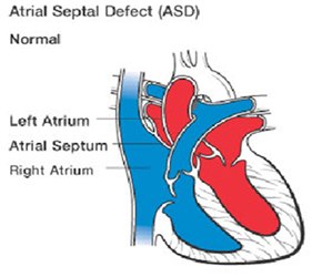
An atrial septal defect allows freshly oxygenated blood to flow from the left upper chamber of the heart (left atrium) into the right upper chamber of the heart (right atrium). There it mixes with deoxygenated blood and is pumped to the lungs, even though it's already refreshed with oxygen. If the atrial septal defect is large, this extra blood volume can overfill the lungs and overwork the heart. If not treated, the right side of the heart eventually enlarges and weakens. In some cases, the blood pressure in your lungs increases as well, leading to pulmonary hypertension. 


Comparing ASD with patent foramen ovale 

The term "atrial septal defect" usually refers to holes in the atria resulting from a lack of atrial septal tissue, rather than those related to a condition called patent foramen ovale (PFO). 

Patent foramen ovale occurs when part of the normal fetal heart circulation fails to close properly at birth. During fetal heart development, a channel (the foramen ovale) is present between the atria to allow blood to bypass the lungs. At birth, once the lungs take over breathing, the hole normally closes. In about one in three people, this opening doesn't close. 


Treatments and drugs for Atrial Septal Defect (ASD) in India

If your child has an atrial septal defect, your doctor may recommend monitoring it for a period of time to see if it closes on its own, while treating any symptoms with medications. 

According to the National Institutes of Health, about half of all atrial septal defects eventually close on their own. About 20 percent close within the first year of life. If a hole hasn't closed early in childhood, it usually won't close on its own. Some small atrial septal defects don't cause any problems and may not need closure, but many require surgery to be corrected. If your child needs treatment, the timing of it depends on your child's condition and whether your child has any other congenital heart defects. 


Surgery of Atrial Septal Defect (ASD) in India

Many doctors recommend repairing an atrial septal defect diagnosed during childhood to prevent complications as an adult. For adults and children, surgery involves plugging or patching the abnormal opening between the atria. 

Doctors can do this through two methods : -


  • Cardiac catheterization : - ASD Closure. A thin tube (catheter) is inserted into a blood vessel in the groin and guided to the heart. Through the catheter, a mesh patch or plug is put into place to close the hole. The heart tissue grows around the mesh, permanently sealing the hole.

  • Open-heart surgery : - This type of surgery is done under general anesthesia and requires the use of a heart-lung machine. Through an incision in the chest, surgeons use patches or stitches to close the hole.

Follow-up care depends on the type of defect and whether other defects are present. For simple atrial septal defects closed during childhood, only occasional follow-up care is needed. For adults, follow-up care may depend on any resulting complications. 

Friday, 30 October 2015

Ventricular Septal Defect in Children - Ventricular Septal Defect Surgery in India

Ventricular Septal Defect(VSD)
A small hole allows little amount of blood to go across the hole. A large hole gets a lot more of blood across the hole. This extra amount of blood coming to the right side of the heart immediately gets transmitted to the lungs from where it goes to the left side of the heart. So, it is really the left side if the heart, the left upper and lower chambers which get to see this extra amount of blood!
VSD when large has to be closed by the 3rd to 6th month of life.  The decision to close the
VSD has to be made well in time to give family enough time to accommodate to the fact that
heart surgery has to be done on their child and that apart from the emotions of the decision
(how so ever safe it might be, parents will take time to decide and accept), there are
financial (or insurance clearance issues also).
  1. VSD with Coarctation
  1. VSD, large with a pneumonia
  1. Large VSD with Severe Malnutrition
What are the ventricles ? What is the Ventricular septum ?
The heart has 2 pumps; pumping blood is the main function of the heart; the 2 pumps are separated by a wall called the ventricular septum. The right pump receives the blue blood which has to go to the lungs for picking up oxygen. This pump has to push blood into its vicinity (heart and lungs being close by within the chest); so the normal pressure in this pump is 30-35 mmHg.
The Left Pump pushes the red blood into the lungs; it has to ensure blood reaches all the parts of the body so the pressure here is equivalent to the blood pressure (upper number of BP i.e. Systolic).
What is a Ventricular septal defect (VSD) ?
A defect in the wall which separates the ventricles is called a VSD. The high pressure in the Left pump as compared to the Right ensures that the blood flows from the left to the right pump (therefore is called a L to R shunt). This implies red (left sided) blood gets into the blue (right sided) blood; this implies that in this condition the patient is not going to be blue.
How common is it?
It is one of the most common defects of the heart. Infact it is the most common defect.
Can it be detected before birth ? 
Yes, it can be detected before and at birth. Often, it will detected on an ultrasound. But on fetal echocardiogram there is a better chance of it being picked up. The way this hole is noticed, is by seeing abnormal flow across the septum. But, during fetal life the pressure between the 2 pumps is the same. So, there is not much flow across the hole and that makes it difficult to pick up.
How does a small hole differ from a large hole?
If this blood amount is large, the left side of the heart gets dilated and volume overloaded. This can be seen on an X-ray as enlarged heart.  This reverses once the extra blood flow stops.
If the amount of blood is large, there will also be an increase in pressure in the right side of heart and in the lungs. While with the Small VSD the volume will be so small the left heart won’t enlarge. And neither will the pressure in the right heart go up.

Is VSD closure an emergency ?
Usually VSD closure is an elective procedure done in a planned manner, but, ocassioanlly
may turn out to be emergency in some circumstances:
A ventricular septal defect usually is diagnosed after a baby is born.

Doctors may choose to monitor and observe children who have ventricular septal defects (VSDs) but don't have symptoms of heart failure. This means regular checkups and tests to see whether the defect closes on its own or gets smaller.
More than half of VSDs eventually close, usually by the time a child is in preschool. Your child's doctor will let you know how often your child should be checked. Checkups may range from once a month to once every 1 or 2 years.
When treatment for a VSD is required, options include extra nutrition and surgery to close the VSD.
The doctor may recommend surgery if your child's VSD:

Some infants who have VSDs don't grow and develop or gain weight as they should. These infants usually:
Doctors usually recommend extra nutrition or special feedings for these infants. These feedings are high-calorie formulas or breast milk supplements that give babies extra nourishment.
In some cases, tube feeding is needed. Food is given through a small tube that's placed through the nose and into the stomach. Tube feeding can add to or take the place of bottle feeding. This treatment usually is temporary because a VSD that causes symptoms will likely need surgery.

Most doctors recommend surgery to close large VSDs that are causing symptoms or haven't closed by the time children are 1 year old. Surgery may be needed earlier if:
Rarely, medium-sized VSDs that are causing enlarged heart chambers are treated with surgery after infancy. However, most VSDs that need surgery are repaired in the first year of life.
Diagnosis
 
  • Shortness of breath,
  • Fast or heavy breathing,
  • Sweating,
  • Tiredness while feeding, or
  • Poor weight gain.
  • Is large
  • Is causing symptoms
  • Is medium-sized and is causing enlarged heart chambers
  • Affects the aortic valve
Extra Nutrition
  • Have large VSDs
  • Are born prematurely
  • Tire easily during feeding
  • The child fails to gain weight
  • Medicines are needed to control the symptoms of heart failure

Sunday, 25 October 2015

Pediatric Cardiac Surgery at World's Class Hospital in India

Heart Surgery in India

Hospitals in India have a success rate of between 98 to 99% for heart surgery in India Heart surgery in India is performed by cardiac surgeons of great experience, in terms of both number of surgeries, diversity of procedures and complexity of heart conditions This experience is built on top of the outstanding academic qualifications of heart surgeons in India.
Sometimes heart surgery in children is required for repairing defects in the heart which a child might have born with (known as congenital heart defects) and heart diseases which he gets after birth, which require surgery. Pediatric Cardiac Surgery deals with operative procedures in the newborn and unborn children and youngsters suffering from cardiac dysfunctions, structural, functional and rhythm-related issues of the heart also.

Pediatric Cardiac Surgery often deals with heart problems in children, the cause of congenital heart diseases which are common heart ailments among new born children and involves deficiencies like structural defects, congenital arrythmias and cardiomyopathies, which result in different kinds of abnormalities related to the heart. Congenital heart disease is a cause of improper growth of the heart or blood vessels before birth.

Pediatric Cardiology Facilities
The Paediatric cardiology department of any hospital offers all non-invasive diagnostic and paediatric cardiac interventional services. The diagnostic modalities are inclusive of foetal echocardiography, transthoracic and transesophageal echocardiography, round the clock Holter, CT and MR angiography as well as diagnostic cardiac catheterisation. Cardiologists, cardiovascular surgeons, anesthesiologists and other specialists work closely together to care for newborn as well as unborn children suffering from cardio-vascular disorders who provide exceptional pre and post operative services.

Pediatric heart surgery often deals with cases of congenital heart disease. Congenital heart disease is a common heart ailment among new born children and involves deficiencies like structural defects, congenital arrythmias and cardiomyopathies. This leads to different kinds of abnormalities related to theheart. Congenital heart disease is a cause of improper growth of the heart or blood vessels before birth. Pediatric heart surgery deals with operative procedures dealing with newborn and unborn children and youngsters suffering from cardiac dysfunctions.
There are a number of Pediatric Cardiology Surgeries such as balloon atrial septostomy, Valvuloplasty/Angioplasty, Patent Ductus Arteriosus, Ventricular Septal Defect and Atrial Septal Defect, Tetralogy of Fallot, Total Anomalous Pulmonary Venous Return, Double Outlet Right Ventricle etc. The major ones are:
Balloon Atrial Septostomy (BAS) : This technique, also known as the Rashkind procedure used for enlarging a hole between the right atrium and the left atrium. It is often used to manage patients with transposition of the great arteries. Here the larger hole improves oxygenation of the blood.
Valvuloplasty/Angioplasty : Valvuloplasty is that process where a small balloon is inserted and inflated for stretching and opening a narrowed (stenosed) heart valve. On the other hand, angioplasty is a non-surgical process which can be used for opening blocked heart arteries.

Patent Ductus Arteriosus : This is a kind of defect in infants where vessel (the ductus arteriosus) fails to close soon after birth which results in abnormal blood flow, between the pulmonary artery and aorta, which takes the blood from the heart to the lungs and to the rest of the body. To treat it, an incision is made under the chest through which tubes are inserted to regulate the flow of blood supply.

Atrial Septal Defect (ASD) and Ventricular Septal Defect (VSD) : Both of these are congenital defects. In ASD, the wall that separates the upper heart chambers (atria) does not close completely. The surgery here involves a placement of an ASD device into the heart through catheters, which is inserted by making a tiny incision in the groin area. While in VSD, happens at the time of birth. It is explained as one or more holes in the wall that separates the right and left ventricle of the heart. In pre-natal stages, the right and left ventricles of a heart are not separate. As the fetus grows, a wall is formed for separating the two ventricles but if the wall does not form completely, a hole remains. This can be treated by inserting cardiac catheterization.

Tetralogy of Fallot (ToF) : Another congenital heart defect, it is a constellation of four abnormalities, which result in insufficient oxygen in the blood. In this case, a surgery is needed for restoring these defects which are caused, main; paid at the time when the infant is young.

The cost of heart surgery in India is perhaps the lowest in the world Some comparisons show that patients in the USA can save 90 per cent of the treatment cost by having their heart surgery in India. The cost of heart treatment in India is also very much cheaper compared with Thailand, Singapore, etc which also attract medical tourists.

India’s hospitals have world-class cardiac catheterisation labs for diagnosis of heart diseases and performance of angiograms and angioplasties They also have state-of-the-art operation theatres that enable safe and successful heart surgery in India.”